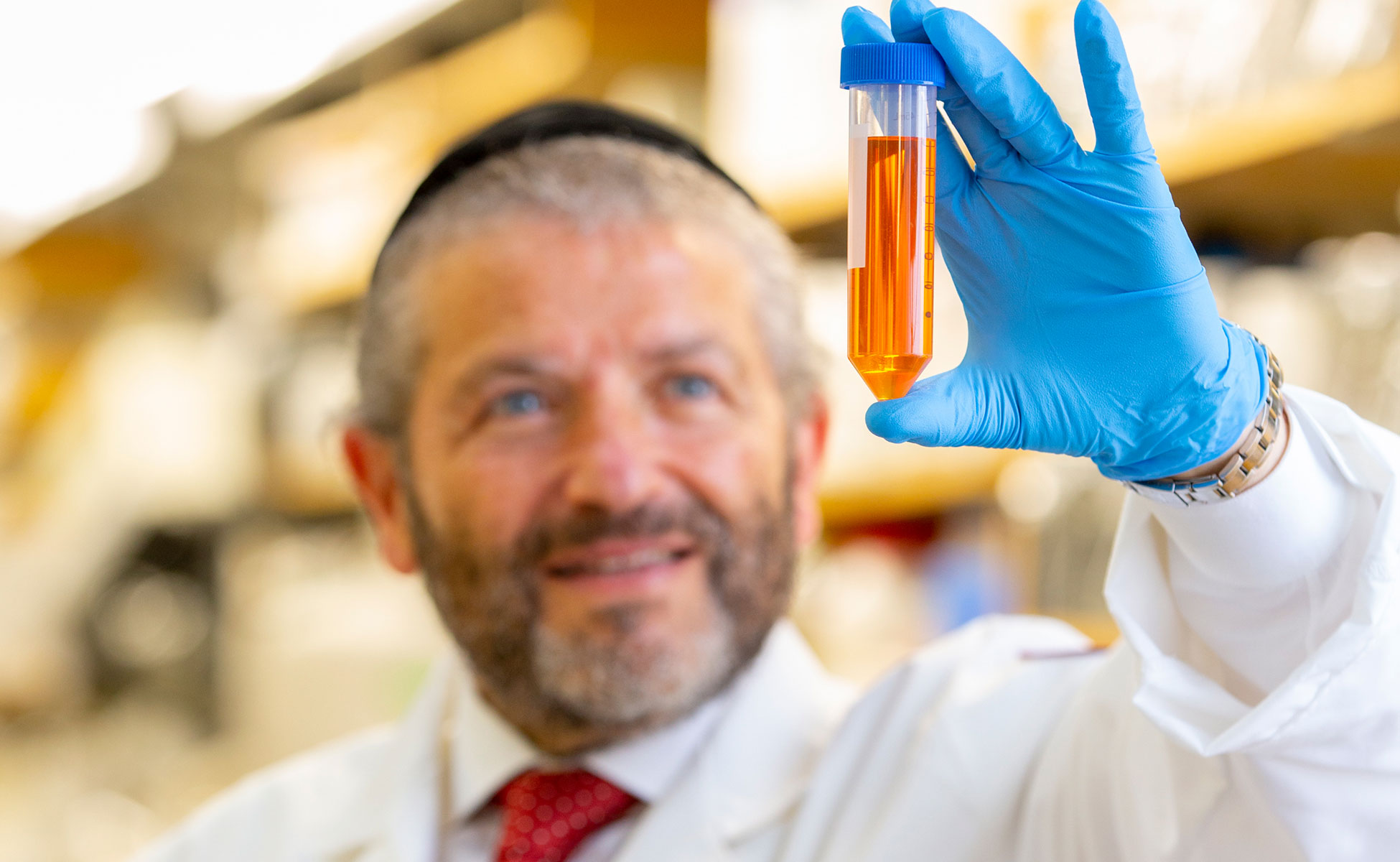
The Histopathology and Imaging Core is located in the Department of Pathology, Microbiology and Immunology, in the Basic Science Building, room 421. The core provides routine histology, special staining, immunohistochemistry, immunoflorescence and cryotomy services, to the research community of NYMC as well as outside entities and institutions. The core has full time personnel responsible for day-to-day operations and training of users on Imaging Core Instrumentation.
The Histology Laboratory accepts fixed wet tissue specimens (in labeled tissue cassettes) as well as frozen tissue samples. The core can provide paraffin and frozen sections. Sections can be prepared on standard or charged slides and stained with either Hematoxylin and Eosin (H&E) or special stains, or left unstained. The lab is semi-automated to provide consistent results and quick turnaround. Our histology protocols strive to maintain the highest possible specimen quality and minimize the possibility of cross contamination.
Equipment
Leica Microdissection System LMD 6500
The Leica Microdissection system LMD 6500 is used for obtaining well-defined starting material for subsequent downstream applications such as genomics, RNA transcript profiling, proteomics and microarrays. With this instrument one can quickly locate a single cell or large group of cells, technically draw and cut and remove specific cells for subsequent molecular analysis. Regardless of whether one uses paraffin-embedded or frozen tissue samples, stained or immuno-labeled slides, the LMD preserves the exact morphologies of both the captured cells as well as the surrounding tissue. This microscope has the following objectives:
- 40x HCX Plan FL 0.6na CORR XT
- 20x HCX Plan FL 0.4na CORR
- 10x HC Plan FL 0.3na
- 5x UVI Microdissection 0.12na
Nikon 90i Eclipse Research microscope
The Nikon 90i Eclipse Research microscope is fully automated for light and fluorescent microscopy. Cubes available are DAPI, GFP (488) and Texas Red (593). Software is available for various morphometric analysis. Objectives available at:
- 4X Plan Fluor
- 10X Plan Fluor
- 20X Plan Fluor40X Plan Apo
- 60X oil Plan Apo
- 100X oil Plan Apo
Zeiss LSM980 plus Airyscan 2 Confocal System
The Zeiss LSM980 plus Airyscan 2 Confocal System is a high-resolution high sensitivity advanced confocal imaging system that is fully equipped for 3D and 4D imaging of live and fixed cells and tissues as well as photobleaching studies. It is located in the Basic Science Building room 245. The system microscope objectives include:
- Super-resolution 63x1.4 NA high resolution objective
- 10x/0.3 NA
- 20x/0.8 NA air objective
- Long Distance 40X/1.2 NA Multi-Immersion objective
Learn more about our Confocal 4D Imaging System.
Other Equipment
- Leica ASP300 tissue processor
- Leica CM1850 cryostat
- Sakura Tissue-Tek TEC-5 tissue embedding console system
Service Request
Investigators who would like to request services from the laboratory should contact us. We will send a requisition form to fill out. The PI is required to sign the request form. The services provided will be based on the order in which it is received.
Histology Services Contact
S M Shafiqul Alam
Core Lab Manager
(914) 594-4862
salam8@nymc.edu
Tetyana Cheairs, M.D., M.S.P.H.
Core Director
(914) 594-3105
tetyana_kobets@nymc.edu
Pathology Consultations Contact
Humayun Islam, M.D., Ph.D.
Chair, Department of Pathology, Microbiology and Immunology
P: (914) 594-4150
F: (914) 594-4163
humayun.islam@wmchealth.org


